Stratipath Breast is the first AI-based prognostic risk profiling tool, regulatory aligned for clinical use in the EU and the UK. The solution analyses cancer tissue, enabling the identification of patients with either an increased, or low, risk of disease progression. It is already in clinical use and validated in large-scale studies, ensuring robust and reliable performance.
AI-based risk stratification enables faster turnaround times for results, provides new information at the point of diagnosis, and can reduce the need for expensive gene expression testing, allowing for wider use and benefit to more patients.

“For patients, shortening the time to a treatment decision can provide immense relief, sparing them weeks of uncertainty and worry.”
Oncologist using Stratipath Breast

Stratipath Breast has been evaluated in independent validation studies and has been used in clinical settings for over a year, proving its reliability in real-world diagnostics.
Stratipath Breast provides a faster and a more cost-effective alternative to gene expression tests. By leveraging AI to analyse routine pathology slides, it can reduce lab time and costs, delivering risk assessments within the hour instead of weeks.


Stratipath Breast analyses digitalised routine HE images of breast cancer to deliver prognostic information associated with a patient’s risk of relapse, providing critical insights to support physicians in making informed treatment decisions.

“Stratipath Breast offers a faster and cheaper alternative to gene expression testing, allowing more patients to have access to precision diagnostics. By using Stratipath Breast, clinicians can diagnose with support from independent prognostic analysis, while reducing laboratory time and costs.”
Johan Hartman
Professor of pathology at Karolinska Institutet, Stockholm and co-founder of Stratipath
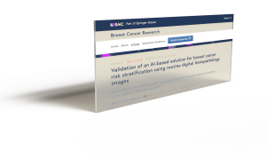
Stratipath Breast builds on academic research previously published in Annals of Oncology, which demonstrated the potential of AI-based risk profiling for breast cancer. Since then, multiple studies have been published, including a large validation study demonstrating the prognostic performance of Stratipath Breast.


“The validation of our earlier findings using the CE-marked product highlights the reliability and effectiveness of our AI-based approach. We are proud that Stratipath Breast is already making a difference for many patients, and we look forward to expanding its use further.”
Johan Hartman
Professor of pathology at Karolinska Institutet, Stockholm and co-founder of Stratipath
• Intended for female patients with invasive breast cancer of all grades, supporting broader prognostic testing.
• As an alternative to gene expression tests.
• Developed and optimised in cohorts including both pre- and postmenopausal patients.
• For use irrespective of the patient’s lymph node status
• For early-stage breast cancer, not to be used on metastatic recurrences

Unilabs, a leading diagnostic services provider in Europe, has announced the implementation of Stratipath Breast, an AI-powered precision diagnostic tool for breast cancer patients, at Capio St Göran’s Hospital in Stockholm.
Stratipath Breast has been successfully implemented in clinical routine at multiple healthcare sites, where it is used daily for risk profiling of breast cancer patients. By integrating seamlessly into existing diagnostic workflows, Stratipath Breast provides healthcare providers with a faster, more affordable alternative to gene expression tests, reducing both time to treatment and overall healthcare costs. Stratipath Breast is CE-IVD marked and registered in the UK, enabling its clinical use in both the EU and the UK.

“Stratipath Breast enables us to provide more patients with high-quality diagnostic insights faster than ever before.”
Eugenia Colón
senior consultant pathologist at Unilabs
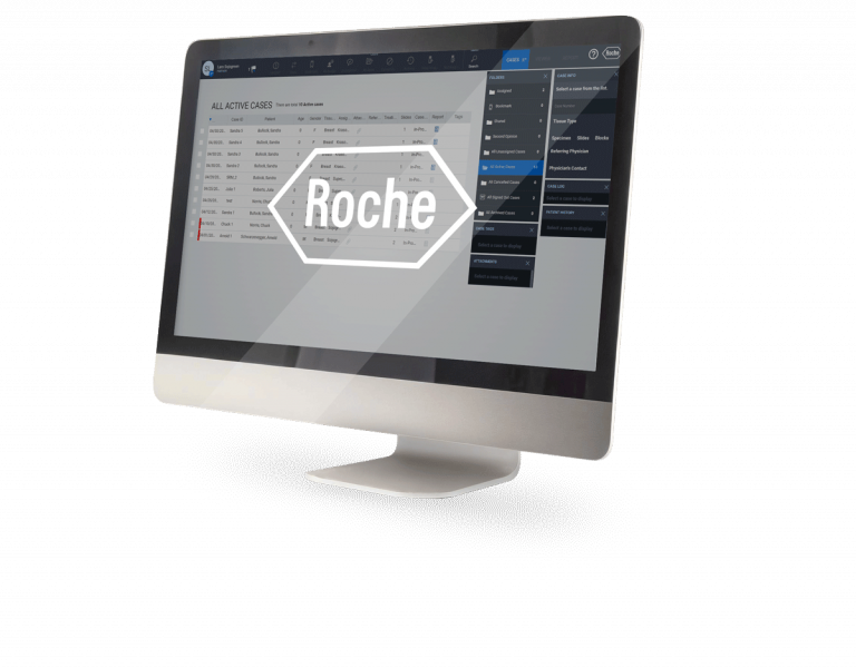
Stratipath Breast provides an optimal workflow through integration with leading digital pathology solutions. It can also be used on its own, via the Stratipath customer portal. Access to Stratipath Breast will be provided as a Software as a Service solution, by a subscription model.
”The answer is there when you need it – ready before the tumour board.”
/ ONCOLOGIST USING STRATIPATH BREAST
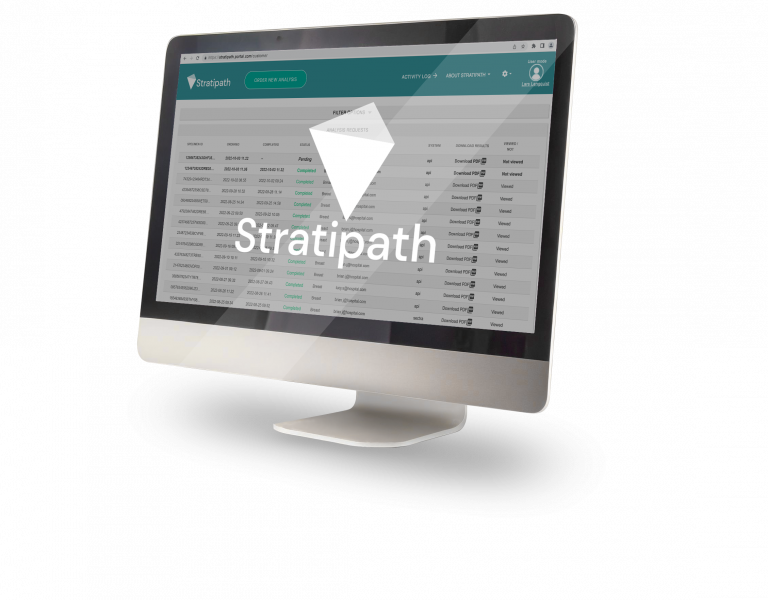
The results from the Stratipath Breast analysis are available as a PDF report, ready to download, share or include in the diagnostic report available for the tumour board.
